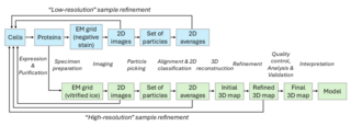
Cryo-electron microscopy (cryoEM) is a powerful technique for studying the structures of biological samples that are rapidly frozen (vitrified) at liquid nitrogen temperatures. It encompasses a range of applications, including single-particle analysis (SPA), cryo-electron tomography (cryoET), 2D electron crystallography, and microcrystal electron diffraction (MicroED).
1. Single particle analysis of purified protein complexes
Cheng Y, Grigorieff N, Penczek PA & Walz, T. A primer to single-particle cryo-electron microscopy. Cell. 2015, 161: 438–449.